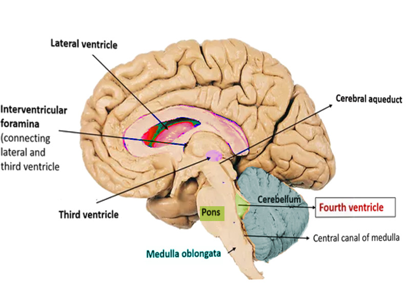

They may also be enlarged in a number of neurological conditions (e.g. The volume of the lateral ventricles is known to increase with age due to cerebral involution. Why does the volume of the lateral ventricle increase? Cerebrospinal fluid, or CSF, is made and stored in the brain’s ventricles. It happens when one or more ventricals, which are normally hollow areas in the brain, have too much cerebrospinal fluid. Have a … read more What does it mean when your brain ventricles are enlarged?Įnlarged ventricles in the brain may be a sign of normal pressure hydrocephalus. This is what it said “Minimal periventricular subcortical scattered gliosis consistent … read more Have had CAT scan and MRI both indicate marked prominence of both lateral ventricles….third a little, fourth normal in size. Ultrasound is the screening modality of choice for initial evaluation 8. While many fetuses with mild ventriculomegaly have a normal outcome, there are also a large number of congenital syndromes associated with enlarged ventricles. Can a fetus with mild ventriculomegaly be normal? The choroid plexus begins to recede posteriorly but remains in close contact with the medial and lateral walls of the bodies and atria of the ventricles While many fetuses with mild ventriculomegaly have a normal outcome, there are also a large number of congenital syndromes associated with enlarged ventricles. What happens to the choroid plexus during ventriculomegaly? The prevalence of asymmetry in lateral ventricle size in those without evidence of underlying etiology has been found to be 5-12% 1-3. This asymmetry of the lateral ventricles (ALV) is an anatomic variant in most cases. The lateral ventricles occasionally show small side to side differences in size on CT or MRI of the brain. Is there asymmetry in the lateral ventricles of the brain? What is a minimally prominent lateral ventricle of? What happens if the lateral ventricle is enlarged?Ī minor self-limiting imbalance in fluid circulation and absorption that becomes compensated. Ventricles develop early in pregnancy and can be seen on a prenatal ultrasound in the second trimester, at about the 15th week.

This can occur when CSF becomes trapped in the spaces, causing them to grow larger. Ventriculomegaly is a condition in which the ventricles appear larger than normal on a prenatal ultrasound.

Mild ventriculomegaly is different from the more severe form of ventriculomegaly, hydrocephalus or “water on the brain”. Ventricles measuring between 10 to15 mm are called “mild” ventriculomegaly. Mild ventriculomegaly means that one or both of the baby’s lateral ventricles are slightly larger than usual. What is mild prominence of the lateral ventricles?


 0 kommentar(er)
